A Giant Mesenchymal Pericardial Cyst: A Rare cause of Hoarseness of Voice, Case Report and Review of Literature-Juniper publishers
JUNIPER PUBLISHERS-OPEN ACCESS INTERNATIONAL JOURNAL OF PULMONARY & RESPIRATORY SCIENCES
Abstract
We report a case of 32 years women who presented to
us with history of shortness of breath and hoarseness of voice for the
last three weeks. Chest x-ray showed wide mediastinum. Computed
tomographic scan of chest (CT) showed large anterior mediastinal cystic
mass. Fiber optic bronchoscopy showed impaired movement of left vocal
cord. A giant cyst was excised through median sternotomy and histology
report revealed, “Mesenchymal pericardial cyst”.
Keywords: Mediastinal cyst; pericardial; Hoarseness of voice; Surgery.
Introduction
Pericardial cysts are exceedingly rare benign lesions
which constitutes 35% of the mediastinal cystic lesions and 7% of
overall mediastinal masses. The reported incidence of pericardial cysts
is 1:1, 00,000, commonly seen in the third or fourth decade of life
equally affecting both genders [1,2]. In 1958 Le Roux reported only
three cases of pericardial cysts after reviewing 3000 chest x-rays. They
are described in medical literature under various names
pleuropericadial cyst, pleural cysts, spring water cysts, pericardial
coelomic cysts [3]. First surgical resection for pericardial cyst was
carried out by Lenox Hill in 1931 [4].
Case
A 32 year old woman presented with two months history
of shortness of breath and recently changes of voice. No history of
cough, appetite or weight loss. Routine blood investigations including
liver and renal panels were normal. Chest x-ray and CT scan of thorax
showed a large cyst mass occupying the anterior mediastinum abutting
with the blood vessels (Figure 1A-1C). Pulmonary function test flow
volume loop showed obstructive pattern extrinsic compression.
Echocardiogram was normal.


Surgical approach was median sternotomy and we found a
giant cystic mass occupying the entire anterior mediastinum. The
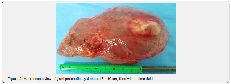
mass was resected in total with careful dissection as shown in
Figure 2. Sternum was closed with wires. Patient was extubated
on table and later on transferred to high dependency unit for
overnight observations. Post-operative recovery was uneventful
and she was discharged home. Five weeks later she check fibro
optic bronchoscopy showed normal movements of vocal cards
and her voice returned back to normal as well .Follow up chest
x-ray and CT of thorax showed mediastinal mass. (Figure 3D-3F)
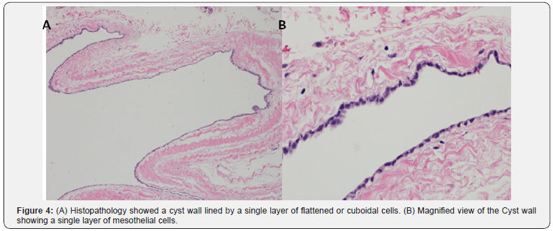
Histopathology report showed benign mesenchymal pericardial
cyst (Figure 4A & 4B).
Discussion
Pericardial cysts are rare benign congenital lesions which
occur due to failure of fusion of mesenchymal lacunae which form
pericardial sac during embryogenesis [5,6]. Acquired pericardial
cysts have been reported due to rheumatic pericarditis,
tuberculosis. Hydatid disease, trauma and cardiac surgery [7].
They are commonly located at right or left cost phrenic angles
70% and 20% respectively, rest of 10% can be anywhere in
the anterior or posterior mediastinum [8]. The best diagnostic
imaging modality is CT scan or MRI scans of thorax although
cardiac computed tomography and cardiac magnetic resonance
imaging provides additional information about its relation with
the heart. Normally these cysts contain clear fluid and diagnosis
is easy but if the fluid protein content is high or infected then
it’s very difficult to differentiate them from other mediastinal
malignant lesions [9,10]. Echocardiography is very useful tool to
delineate the lesions located close to heart borders. Pericardial
cysts are usually detected as incidental finding during chest
imaging and majority of them are asymptomatic and are treated
conservatively. Pericardial cysts can be detected prenatally after
14th week of gestation and can regress spontaneously [11]. The
symptomatic pericardial cysts are resected surgically as they can
cause a lot of cardiac and non-cardiac complications due to their
mass effect or by direct invasion of adjacent structures [12-16]
(Table 1).

Feign et al. [17] reported thirty two patients out of 82
cases were symptomatic and they presented with chest pain,
dyspnea, cough paroxysmal atrial tachycardia, pneumothorax,
and hemoptysis. Unverferth et al. [18] reported series of twelve
cases of pericardial cysts only two of them were symptomatic.
Menconi et al. [19] reported three out of five cases with similar
symptoms all of them were treated by surgical resection of cysts.
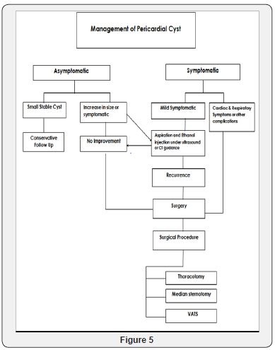
Management of pericardial cyst is three dimensional,
conservative, aspiration and surgical resection Figure 5.

The European society of cardiology task force for management
of pericardial diseases recommended percutaneous aspiration of
pericardial cysts and ethanol sclerosis as first line of treatment
[20].
Conclusion
We present a case of young women who presented with
hoarseness of voice and shortness of breath due to giant
pericardial cyst causing the compression of airway and left
recurrent laryngeal nerve.
Pericardial cyst was excised in
total with excellent post-operative result. Patient dramatically
improved after surgery and follow up bronchoscopy after five
weeks showed normal movements of left vocal card and patient
regained her normal voice.
To know more about Open Access International
Journal of Pulmonary & Respiratory Sciences please click on: https://juniperpublishers.com/ijoprs/index.php
To know more about Open access Journals
Publishers please click on : Juniper Publishers

Comments
Post a Comment