Candida Albicans Bone Rib Osteomyelitis due to Previous Systemic Candidiasis-Juniper publishers
JUNIPER PUBLISHERS-OPEN ACCESS INTERNATIONAL JOURNAL OF PULMONARY & RESPIRATORY SCIENCES
Abstract
Candida albicans osteomyelitis causes significant
morbidity if not recognized early. Due to its rarity and chronic course
symptoms may persist for months before diagnosis is suspected.
Pathogenesis is related to previous candidemia and costochondral joints
represent an usual localization.
Keywords: Candida albicans; Osteomyelitis; Costochondral junction; Candidemia
Abbrevations: CA: Candida Albicans; ICU: Intensive Care Unit; WBC: White Blood Cells; CT: Computed Tomography
Introduction
A 45-years old man referred to our unit for a painful
and swelling mass in the right inferior parasternal joint. The mass
appeared several months before, without sings of resolution despite
empiric antibiotical therapy. Remarkable in patient’s medical history
was that on July 2017 a routine coronary angiography caused total
dissection of his right coronary artery up to the main coronary sinus,
treated by emergency surgical repair and coronary by-pass. After surgery
the patient developed a bilateral pneumonia with excavated abscess in
the lung after a long invasive ventilation; multi drugs resistant
Acinetobacter was isolated in bronchial lavage with Klebsiella
Pneumoniae plus Candida Albicans isolated in blood cultures. The CT
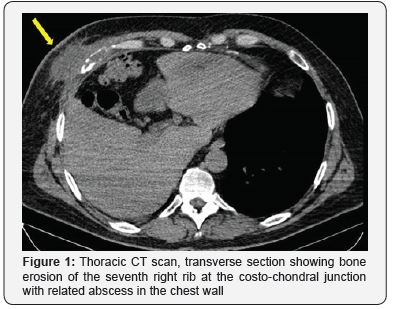
study showed an area of bone erosion in the seventh right rib at the
costo-chondral junction with related abscess in the chest wall up to the
skin. The patient was treated surgically by en-block resection of the
involved bone and chest wall abscess, with reconstruction of the chest
wall made by polyglactin knitted (Vycril®) associated with a prolonged
anti-mycotic therapy.
Conclusion
Post-surgical C.A. osteomyelitis is more often due to
direct insemination and it is subsequent to C.A. hematogenous spread in
people harboring predisposing factors like recent major surgery,
immunosupression and prolonged ICU stay.
Case Description
On January 2018 a 45 years old man referred to our
unit for a painful and swelling mass in the right inferior parasternal
joint. The mass appeared several months before, without sings of
resolution despite empiric antibiotical therapy. Remarkable in patient’s
medical history was that on July 2017 a routine coronary angiography
caused total dissection of his right coronary artery up to the main
coronary sinus, treated by emergency surgical repair and coronary
by-pass. After surgery the patient had a prolonged ICU stay with
prolonged mechanical
ventilation developinga bilateral pneumonia with excavated
abscess in the lung. Multi drugs resistant Acinetobacterwas
isolated in bronchial lavage and Klebsiella Pneumoniae plus
Candida Albicans were isolated in blood cultures.


A various range of antibiotical and antimycothic therapy was
performed, and finally the patient was discharged from ICU in
September and he finally was discharged in November 2017.
After ICU discharge he presented the same swelling and painful
mass in lower right parasternal region as we observed; previous
median sternotomy presented well consolidated, far and without
any involvement in the abscess. On admission he had no fever
and normal WBC count; the CT study showed an area of bone
erosion in the seventh right rib at the costo-chondral junction
with related abscess in the chest wall up to the skin (Figure 1
& 2) and confirmed no involvement of the sternotomy. He was
treated surgically by en-block resection of the involved bone and
chest wall abscess, with reconstruction of the chest wall made
by polyglactin knitted (Vycril®, Ethicon) absorbable mesh fixed
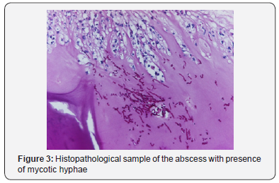
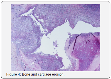
on the near ribs.Histopathology samples confirm abscess and
bone erosion with presence of mycotic hyphae inside (Figure 3
& 4); culture samples from bone were positive for C.A.Promptly
specific medical therapy against C.A. was started and planned for
a six months lasting period. In the eleventh post-operative day
he was discharged at home with oral therapy with fluconazole
600 mg per day for six months.
Discussion
The first reports about costo-chondral involvement in
systemic candidiasis dates the eighties and intravenous
heroin addicts were involved [1]. Typically, the lack of hygiene
in their intravenous injection causes C.A. hematogenous
dissemination and subsequent osteoarticular involvement
mainly in costo-chondral joint [2]. C.A. hematogenous spread
and immunosuppression plays an important role in the
pathogenesis for non-traumatic patients or without direct
local insemination.C.A. osteomyelitis may be divided in
proven osteomyelitis (i.e. compatible clinical characteristics,
radiological appearance and positive cultures) or only probable
(i.e. positive cultures from bone).Due to its rarity and chronic
course symptoms persists for weeks or months before diagnosis
is suspected. A swelling mass with pain and edema represents
the usual presentation; high fever and important increase of
inflammatory markers is unusual.Diagnosis confirmation is made
on CT appearance and, as treatment by surgical debridement
or resection is mandatory, on cultures and histopathology of
surgical specimens.In 2009 IDSA guidelines recommended
antifungal therapy lasting at least six months [6] and state that
premature discontinuation in medical therapy represents the
leading cause of disease relapse [1,6].
Conclusion
Postsurgical C.A. osteomyelitis is uncommon and more often
due to direct insemination1; more rarely, like in our report, it
can be subsequent to C.A. hematogenous spread. This kind of
pathogenesis with elective localization in the costo-chondral
junction was first reported in the eighties in intravenous heroin
addict [2]. In more recent times it is anedottically reported in
people harboring predisposing factors like recent major surgery,
immunosuppression and prolonged ICU stay [1,3-5].
Lesson learned
Due to its rarity C.A. osteomyelitis is not suspected and the
diagnosis is done after weeks or months of pain and inadequate antimicrobial therapy, causing significant morbidity.Prolonged
medical therapy is not enough to solve the problem and surgical
debridement is needed. This can also confirm the diagnosis and
drive the therapy by culture samples.Despite aggressive surgery,
prolonged anti-mycotic therapy for at least six months is needed
and early discontinuation of the therapy represents the leading
cause of infection relapse [1,6,7]. Reconstructing material
interposition represents a predisposing factor for persistent
infection, so the prompt use of metal materials for bone
reconstruction should be avoided. State the need of prolonged
medical therapy we decided for a temporary reconstruction of
the chest wall made by absorbable material, that won’t be in site
at the end of the six months therapy. If bone reconstruction made
by metal material is needed for aesthetic or functional problems
it can be deemed at the time of local sterilization.
Acknowledgment
Drs. Ratti and Fronti kindly provided important indications
for treatment protocols and diagnosis of the CA infection
Dr. Zangrandi kindly provided the histopathological sample
images for publication
Conflicts of interest
The authors have no conflicts of interest to disclose
To know more about Open Access International
Journal of Pulmonary & Respiratory Sciences please click on: https://juniperpublishers.com/ijoprs/index.php
To know more about Open access Journals
Publishers please click on : Juniper Publishers

Comments
Post a Comment