A Case of Systemic Sclerosis with ILD and Pulmonary Tuberculosis - A Rare Case Report-Juniper publishers
JUNIPER PUBLISHERS-OPEN ACCESS INTERNATIONAL JOURNAL OF PULMONARY & RESPIRATORY SCIENCES
Introduction
Systemic scleroderma is an autoimmune disorder that
affects the skin and internal organs. Autoimmune disorders occur when
the immune system malfunctions and attacks the body’s own tissues and
organs. The word “scleroderma” means hard skin in Greek, and the
condition is characterized by the buildup of scar tissue (fibrosis) in
the skin and other organs. The condition is also called systemic
sclerosis because the fibrosis can affect organs other than the skin.
Fibrosis is due to the excess production of a tough protein called
collagen, which normally strengthens and supports connective tissues
throughout the body. The classification of SSC is subdivided based on
the extent of skin involvement into Diffuse Cutaneous Sclerosis (DCSSC),
Limited Cutaneous Sclerosis (LCSSC) or SSC sine scleroderma [1]. Early
sign of systemic scleroderma is puffy or swollen hands before thickening
and hardening of the skin due to fibrosis. Fibrosis can also affect
internal organs and can lead to impairment or failure of the affected
organs. The most commonly affected organs are the esophagus, heart,
lungs, and kidneys. Pulmonary involvement is common in patients with
Systemic Sclerosis (SSC), and this leads to substantial morbidity and
mortality.
While virtually any organ system may be involved in
the disease process, fibrotic and vascular pulmonary manifestations of
SSC, including Interstitial Lung Disease (ILD) and Pulmonary
Hypertension (PH), are the leading cause of death. In scleroderma, the
two most common types of direct pulmonary involvement are ILD and PAH,
which together account for 60%
of SSC-related deaths [2]. ILD is common in scleroderma. In early
autopsy studies, up to 100% of patients were found to have parenchymal
involvement [3]. As many as 90% of patients will have interstitial
abnormalities on High-Resolution Computed Tomography (HRCT) [4] and
40-75% will have changes in Pulmonary Function Tests (PFTs) [5].
Parenchymal lung involvement often appears early after the diagnosis of
SSC, with 25% of patients developing clinically significant lung
disease within 3yrs as defined by physiological,
radiographic or Bronchoalveolar Lavage (BAL) abnormalities. Disrupted
immunity from the disease or associated medication may render such
patients subject to tuberculosis infection. Infections are also known to
induce the development of auto antibodies. A rare case of SSC,
simultaneous diagnosis of ILD and pulmonary tuberculosis is being
reported here.
Case Report
A 58year old female presented with complaints of
breathlessness on exertion (MMRC grade II), cough with expectoration,
fever, loss of appetite, difficulty in swallowing and tightening of skin
around the mouth and skin thickening bilaterally in hands. There was no
history of chest pain, hemoptysis, and wheeze. There was no
occupational exposure to chemicals, dust and smoke.
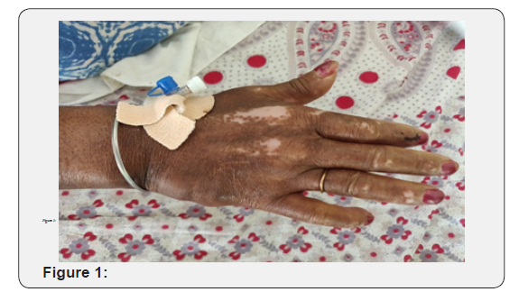
On examination, patient was moderate built. Patient
had features of salt and pepper appearance, mask like face, pinched
nose, facial melanosis, telangiectasia over the cheeks,sclerodactyly and
resorption of digits (Figure 1) diagnostic
of scleroderma. There was no pallor, cyanosis, clubbing,
lymphadenopathy or pedal edema. Pulse was 110/min and BP:
110/70mmhg. Respiratory examination revealed bilateral end
inspiratory crackles of Velcro type and coarse crept over left
suprascapular area. Other systems were normal.

Provisional diagnosis of systemic sclerosis with interstitial
lung disease was made. Due to financial limitation and she was
fitting into clinical ACR criteria [6] for diagnosis, ANA alone
was sent as a part of autoimmune antibody profile which was
positive, value is 5.18 (positive:>1.2), contributing towards the
diagnosis.
Further serological investigations revealed anemia with
Hb of 8.1gm%, elevated leukocyte count with70% polymorphs
and elevated ESR-55mm. Renal and liver functions were within
normal limits. HIV serology was Non-reactive
Chest X-ray (Figure 2) revealed thick walled cavity in the
left upper lobe and increased reticular opacities bilaterally. In
view of thick walled cavity, secondary infections due to bacterial,
fungal and tuberculous organisms were considered. Her sputum
examination for AFB came out to be 3+ and MTB is detected on
CBNAAT the gram stain, cultures revealed no growth.

The skin biopsy was done and histopathlogy report shows
epidermal thickening beneath which bundles of thick collagen
are seen along with adenexal structures and theses features
suggestive of sytemic sclerosis.
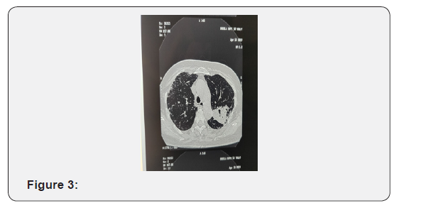
HRCT chest (Figure 3) showed a Soft tissue density lesion
with Fibro-cavitory lesion in the Apico-posterior Segment of Left
Upper Lobe-Likely collapse or consolidation with honeycombing
and Interstitial Thickening involving Interlobular, Centrilobular
and peribronchovascular Intertitium involving both lungs fields
and calcified lymph nodes at bilateral Hilar region likely ILD. So,
she had features of both progressive ILD and active pulmonary
tuberculosis. Patient was initiated on anti tuberculous drugs as
per RNTCP Guidelines and treatment for ILD after 1month of
antituberculous treatment.

Systemic sclerosis is a multisystem disease involving skin,
lungs, kidneys, heart, GI tract and skeletal muscles. Incidence is
three times more in women than in men with the peak incidence
between ages 20-60. Pulmonary involvement in the form of
interstitial lung disease is common in patients with Systemic
Sclerosis (SSC).
Systemic Sclerosis (SSC) has the highest cause-specific
mortality of all of the connective tissue diseases. Although SSC
often affects multiple organ systems, pulmonary involvement,
and in particular Interstitial Lung Disease (ILD), is the leading
cause of death [7]. ILD is present on High-Resolution Computed
Tomography (HRCT) in 55% of patients with SSC on initial
evaluation, but the prevalence is higher (96%) among patients
with abnormal Pulmonary Function Test (PFT) results.
HRCT is the standard method for the noninvasive diagnosis
of SSC-ILD and can detect mild abnormalities. The true incidence
of HRCT abnormalities is difficult to determine, but the majority
of patients (55-84%) will have disease and the extent is generally
limited with an average of 13% of the parenchymal involved [8].
The most common form of ILD appreciated in SSC is
Nonspecific Interstitial Pneumonitis (NSIP); however, other
histopathological manifestations exist, including usual
interstitial pneumonitis, organizing pneumonia, and diffuse
alveolar damage [9]. Other modalities to confirm the diagnosis
are PFT, BAL Cellular profile, Lung Biopsy.
The treatment of SSC-ILD is limited to targeting inflammatory
pathways with corticosteroids or other immunosuppressive
therapy. This therapeutic approach is largely empirical and
parallels strategies historically used in treating idiopathic
pulmonary fibrosis and related disorders. Cyclophosphamide is
currently the most studied immunosuppressive therapy in SSCILD However, the benefit of cyclophosphamide for this disease
is tempered by its complex adverse event profile. More recent
studies have demonstrated the effectiveness of Mycophenolate
systemic sclerosis related ILD, including Scleroderma Lung
study. Tashkin et al. [10] later compared treatment with
Mycophenolate mofetil for 2-years versus cyclophosphamide for
1year. While they noted significant improvement in lung function,
they were unable to confirm greater efficacy at 24 months with
Mycophenolate mofetil despite its superior tolerability and
toxicity profile [10].
Patients with autoimmune diseases are known to develop
infections like tuberculosis either due to the disease activity or
secondary to the immunosuppressive therapy. Tuberculosis per
se is known to induce the development of autoantibodies which
in turn stimulate the manifestation of autoimmune diseases
[11]. Sreeram V Ramagopalan et al. [12] analyzed a database
of statistical records of patients in England (1999 to 2011) and
found a significant association of tuberculosis and autoimmune
diseases. High levels of risk for tuberculosis was found in
diseases like Addison’s disease, SLE, polymyositis and patients
with scleroderma had a relative risk of 6.1 (95% CI 4.4 to 8.2).
He also looked at the incidence of immune diseases after 5 years
of first occurrence of tuberculosis and found significant increase
in Addison’s disease, Sjogren’s and SLE. The possible mechanism
for this association was tuberculosis being infective trigger
acting by molecular mimicry, bystander activation or acting as
an adjuvant.
Pradhan et al did a screening of auto antibodies in
tuberculosis endemic areas and concluded a possible role of
mycobacterial infection triggering autoimmunity. Importance of
screening all tuberculosis patients for autoantibody profile and
follow up for autoimmune related symptoms was highlighted
[11].
In a similar previous case report by Agarwal et al. [13] in
1977 pulmonary tuberculosis was diagnosed in a known case
of scleroderma who has taken systemic steroids for six months
duration. In that case there was a possibility of Tuberculosis
because of immunosuppression secondary to steroid therapy.
Shachor et al. [14] suggests that a diffusely damaged lung
increases the susceptibility to tuberculosis or activation of
dormant tuberculosis [14].
Another study done by Subramanian et al. [15] highlights
the association of tuberculosis and autoimmune diseases [15].
The present case was diagnosed to have interstitial lung disease
and pulmonary tuberculosis concurrently and had characteristic
features of systemic sclerosis.
To know more about Open Access International
Journal of Pulmonary & Respiratory Sciences please click on: https://juniperpublishers.com/ijoprs/index.php

Comments
Post a Comment