Ultrasonography Assessment of Diaphragm in Asthmatic Children and Effects of Diaphragm Strengthening Exercise on Angiogenin Level and Pulmonary Functions-Juniper publishers
JUNIPER PUBLISHERS-OPEN
ACCESS INTERNATIONAL JOURNAL OF PULMONARY & RESPIRATORY SCIENCES
Abstract
Objective: The study aimed at
assessment of diaphragmatic thickness, excursion and fatigue in
asthmatic children with various clinical and functional grading and also
to assess the effect of a specific training program for the diaphragm
“diaphragm strengthening exercise” on pulmonary functions and diaphragm
ultrasonography.
Methods: It included 45 asthmatic
children and 12 healthy children as control group. For all cases,
assessment of clinical severity and control according to GINA guidelines
(2015), spirometric pulmonary functions and serum angiogenin level as
an inflammatory marker were done. sEMG was measured before and after
maximum voluntary ventilation (MVV) maneuver to determine the extent of
diaphragm fatigue. Diaphragm was assessed by ultrasonography for
thickness and excursion. The patients started a special program of
“Diaphragm strengthening exercise” by abdominal weights” for 10-12
weeks, twice per week, then they were re-assessed by spirometric
pulmonary functions and diaphragm ultrasonography.
Results: This study showed a
statistical significant decrease in thickness of diaphragm in asthmatic
children (9.28±1.88mm) Vs(10.4±1.31mm in control) and decreased
excursion of diaphragm(11.06±3.82 mm) Vs (12.05±2.23mm in control)
(p<0.05) with marked impairment of diaphragmatic excursion in
uncontrolled asthmatics compared to both controlled and partly
controlled patients. Serum Angiogenin level was significantly higher in
asthmatics and it was inversely correlated to FEV1 and to diaphragmatic
thickness and excursion. sEMG recording parameters of diaphragm,
including amplitude and frequency were significantly decreased after
maximal voluntary ventilation maneuver (decrease of 18% and 12% from
baseline, respectively), which indicated diaphragmatic fatigue.
Reassessment of asthmatic children after short term exercise training
for the diaphragm showed significant increase in spirometric pulmonary
functions, significant increase in diaphragmatic thickness and
diaphragmatic excursion compared to pre-exercise measurements.
Conclusion: diaphragmatic thickness
and excursion are affected in asthmatics and it is correlated to
pulmonary functions and to inflammatory markers. Diaphragmatic training
programs may be of value in improving symptoms and severity of patients
with asthma through its effect on diaphragmatic thickness and excursion.
Introduction
The abnormality of respiratory muscle functions in
patients with respiratory diseases is multifaceted. Diaphragm is the
inspiratory muscle most shortened during
hyperinflation which is an inevi Table consequence of severe obstructive
airway diseases [1]. In asthmatic patients, inspiratory muscles are,
forced with an increased load. Air flow obstruction increases resistive
work and creates a threshold load that needs to be overcomed with each
breath. An important
goal in managing asthmatic patients should be the reduction of
symptoms especially dyspnea. As respiratory muscle function is
frequently compromised in those patients and it may contribute
to sensation of breathlessness, assessment of their functions are
of clinical importance [2].
Several mechanisms contribute to imbalance between
respiratory load and capacity with consequent respiratory
muscle dysfunction [3]. Although the load on the respiratory
system is well-known to be increased in acute asthma,
ventilatory failure also might result from the failure of the
respiratory muscle pump to generate an adequate negative
intrathoracic pressure. Hyperinflation that accompanies airway
diseases interferes with the ability of the respiratory muscles to
generate subatmospheric pressure and it increases the load on
the respiratory muscles [4].
Different inflammatory markers in asthma can decrease
diaphragmatic contractility by inducing production of reactive
oxygen species which can cause oxidative damage to the
regulatory proteins of sarcoplasma and myofilaments [5].
Angiogenin is an inflammatory marker thought to contribute to
irreversible airway obstruction via formation of new vessels and
the remodeling of the existing vessels [6]. Angiogenin plays a
role in a number of vasculo-proliferative pathologic conditions.
It has been implicated as a mitogen for vascular endothelial cells,
an immune modulator with suppressive effects on polymorph
nuclear leukocytes, an activator of certain protease cascades, as
well as an adhesion molecule [7].
Short term exercise training for the diaphragm has been found
to improve physical fitness and to result in a decrease in number
of asthma attacks, emergency room visits, hospitalization and
days of absence from school. Also a significant improvement in
lung function values and in asthma symptoms may occur [8].
Aim of the work
The aim of this work was to study the changes in diaphragm
as the main inspiratory muscle in asthmatic children with
various clinical and functional severity, also to correlate these
changes with asthma control, spirometric pulmonary functions,
and angiogenin level as an inflammatory marker. The effect
of a specific training program for the diaphragm “diaphragm
strengthening exercise” on pulmonary functions and diaphragm
ultrasonography was also evaluated.
Patients and Methods
This study was conducted on 45 asthmatic children. They
were recruited from the Pediatric Chest Clinic, Ain Shams
University Hospital (23 male and 22 female children); their
mean age was 9.44 ±2.71 years and mean duration of illness was
6.1±3.08 years. Diagnosis of asthma was made according to the
criteria approved by American Thoracic Society [9]. Following
Global Initiative for Asthma classification [10], asthmatics were
classified according to control of asthma into:
Controlled patients (n = 20), 10 males and 10 females, their
ages ranged from 5-15 years with a mean of 9.37 ± 3.57years.
Partly controlled patients (n = 15), 9 males and 6 females,
their ages ranged from 7-15 years with a mean of 10.6 ± 2.79
years.
Uncontrolled patients (n = 10) 4 males and 6 females, their
ages ranged from 10-15 years with a mean of 12.4 ± 1.95 years.
All children were in a Table 1 phase of the disease and did not
suffer from respiratory infections for at least 1 month before the
measurements. Asthmatic children used inhaled corticosteroids
200 to 400 μg twice a day and bronchodilator therapy on
demand. None of the cases were on oral corticosteroids.
Patients with other systemic diseases or who were unable to
perform reproducible lung function maneuvers were excluded
from the study. Twelve age and sex matched healthy children
were considered as a control group.
All studied children completed the following:
- Full history taking and thorough clinical examination.
- Chest radiography (poster anterior).
- Pulmonary function testing using spirometry (Med Graphics 1070 series 2E/1085; Medical Graphics; St. Paul, MN). The measured parameters are:
- FVC (liter) :forced vital capacity .
- FEV1 (liter): Forced expiratory volume in the first second.
- -PEF(liter):peak expiratory flow
- FEF25-75% (liter/second):Forced expiratory flow rate over 25-75% part of FVC.
- Maximal Voluntary Ventilation (MVV)measurement When the MVV was measured, the patients were asked to sit up very straight and make sure nothing was restricting chest movement or airflow. The subjects began the test by breathing normally through the mouthpiece, followed by breathing as deeply (recommended depth: 1/2–3/4 of the patient’s vital capacity) and rapidly (recommended rate: 70 breaths/min) as possible for 15 seconds At the end of the measurement interval, they were told to resume normal breathing and the mouthpiece was removed [9].
- Surface EMG recordings for diaphragm [2] sEMG was measured before and after MVV maneuver to determine the extent of diaphragm fatigue. Surface recordings of the right costal diaphragmatic EMG activity were obtained by using pairs of skin-taped silver/silver chloride electrodes filled with conductive paste. The electrodes were placed in the seventh or eighth intercostal space on the right side of the body at the mid-clavicular line.A ground electrode was placed on the sternum. The distance between the two electrodes of a given pair was kept to a minimum, never exceeding 2cm, and care was taken to place the electrodes in the same orientation as the muscle fibers. Once the electrodes were positioned and a clear EMG signal was confirmed (by a deep inspiration), the electrodes were fixed in place. The influence of the ECG on the EMG signal was minimized by recording from the right side of the body and measurements were done from the segments between QRS complexes. The following parameters were measured during sEMG:
- Human Angiogenin in the serum This was performed using a Quantikine ELISA kit (R&D Systems; Minneapolis, MN). This kit is used for the quantitative determination of human angiogenin. A monoclonal antibody specific for angiogenin has been percolated onto a micro plate. After washing away any unbound substances, an enzyme-linked polyclonal antibody specific for angiogenin is added to the wells. A substrate solution is added to the wells and color develops in proportion to the amount of angiogenin bound. The intensity of color is measured [11].
- Root mean square (RMS) (μV).
- Median frequency of the EMG power spectrum (Hz).
Both parameters used to diagnose fatigue of diaphragm.
Ultrasonographic assessment of diaphragmatic thickness
by high frequency probes (5-7.5M HZ) to observe diaphragm
at zone of apposition which is the part of diaphragm in contact
with lateral chest wall below the inferior border of costophrenic
angle. The transducer is aligned parallel to midline, then a
picture of liver and diaphragm is taken in full inspiration and
in full expiration from same point. A line is drawn between the
angle of the picture and the dome of diaphragm in both sides in
full inspiration and full expiration. The difference between both
points is considered the diaphragmatic excursion [12].
The patients started “Diaphragm strengthening exercise” by
abdominal weights” for 10-12 weeks, twice per week, then they
were re-assessed by Pulmonary function tests and diaphragm
ultrasound.
Diaphragm strengthening exercise
This exercise was done through abdominal weights in
Crock-lying position. The abdominal weights are sand weights
connected to the upper abdomen with adhesive straps to be
applied firmly, their weights were graduated from half kilogram
till three kilograms according to patient’s ability. The exercise
was applied as 2 sessions per week for about 3 months each
session takes about 30 minutes while the patient asked to inspire
deeply 3 times each minute.
Statistical analysis
A standard computer program (SPSS for Windows, Release
10.0; SPSS; Chicago, IL) was used for data entry and analysis. All
numeric variables were expressed as mean ± SD. Comparison of
different variables in various groups was done using Studentt-
test and Mann Whitney test for normal and nonparametric
variables, respectively. Comparisons of multiple subgroups were
done using analysis of variance and Kruskall-Wallis tests for
normal and nonparametric variables, respectively. Pearson and
Spearman correlation test were used for correlating normal and
nonparametric variables, respectively. For all tests, p < 0.05 was
considered significant.
Result
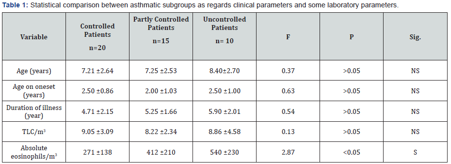
The results of this work were shown in Tables 1-8 and
Figures 1-5. There was a statistical significant difference
between asthmatic subgroups (controlled, partly controlled
and uncontrolled cases) as regards the absolute eosinophilic
count, but there is no statistical significant difference between
them regarding the clinical parameters and TLC/m3 (Table 1).
There was a statistical significant difference between the three
subgroups as regards spirometric pulmonary functions tests
with lower values in uncontrolled patients (Table 2).

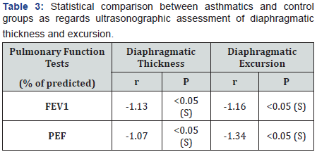
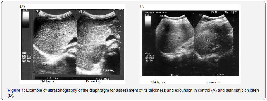
There was a statistical significant lower values of
diaphragmatic thickness and excursion in asthmatics compared
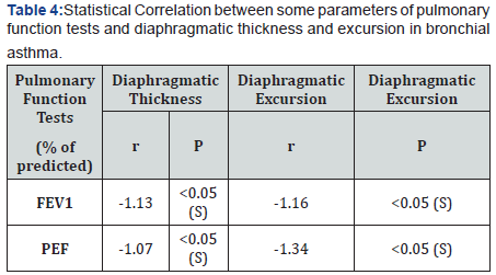
to control. (Table 3 and Figure 1). There was a statistical
significant positive correlation between pulmonary functions
and diaphragmatic thickness and excursion in asthmatics
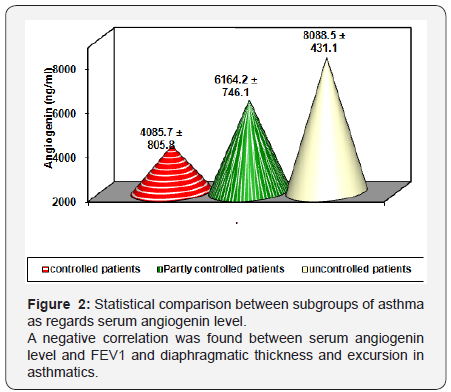
(Table 4). Angiogenin level was significantly higher in asthmatic
children compared to control and was significantly higher in
uncontrolled cases compared to controlled and partly controlled
cases (Table 5 and Figure 2).
sEMG recording parameters of diaphragm, including amplitude
and frequency, were significantly decreased after maximum
voluntary ventilation maneuvers (decrease of 18% and 12%
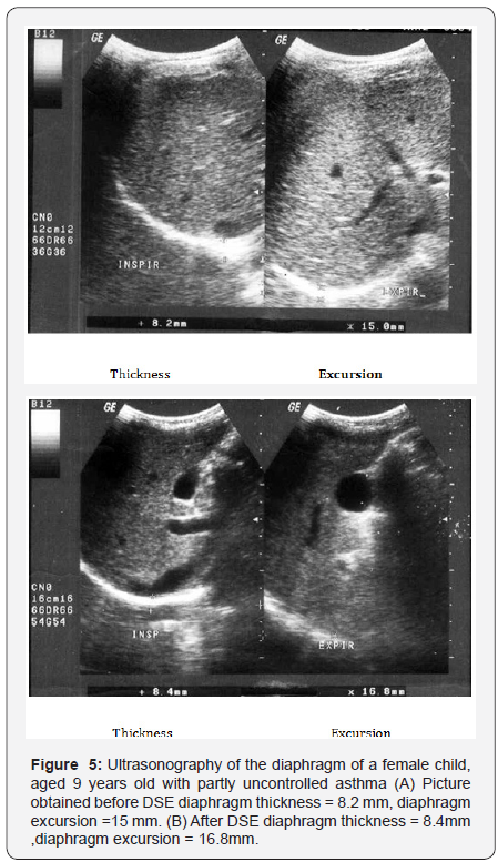
from baseline, respectively), P<0.05 (Table 6). After Diaphragm
strengthening exercise (DSE), a statistical significant higher
values of FEV1, FVC, FEV/FVC, PEF, FEF25-75% and MVV were
detected compared to pre-exercise levels (Table 7). There was
an increase in both thickness and excursion after Diaphragm
strengthening exercise in asthmatic patients (Table 8 and Figure
5).


This study revealed a statistical significant decreased
thickness of the diaphragm in asthmatic patients (9.28±1.88mm)
compared to control group (10.4±1.31mm) (P<0.05), and less diaphragmatic excursion “motion” in asthmatics
(11.06±3.82mm) compared to control (12.05±2.23mm)
(p<0.05). This was in accordance with Targhette [12] who
evaluated diaphragmatic excursion and thickness in asthmatic
children by ultrasonography. They revealed a statistical
significant decreased thickness and less excursion of diaphragm
in asthmatics compared to control. De Bruin [13] recorded
decreased thickness of diaphragm in patients with obstructive
airway disease when assessed by ultrasound. They explained
this finding by a significant increase in the number of type I
fibers (slow twitch) which has the smallest cross sectional area
and decrease in type II (fast twitch) fibers which has the largest
cross sectional area. This fast to slow twitch fiber transition in
the diaphragm can be regarded as an advantageous adaptation
as it will attenuate fatigability of the diaphragm.
Several studies have been reported on the effect of airway
obstruction on diaphragmatic functions [14-20]. However,
hyperinflation was proved by many workers to be the main
underlying etiology. Hyperinflation forces the diaphragm to
operate in an inefficient way for its force-length relationship.
This will decrease the ability of the diaphragm to generate
negative pressure during inspiration. Also hyperinflationcauses flattening of the diaphragm which in turn places it in
a serious mechanical disadvantage being curved upward, so
decreased excursion [12]. Hyperinflation also leads to loss of
axial direction of diaphragmatic fibers and become directed
medially or inwards [4]. Also as the contractile force increase
in order to develop the inspiratory pressure necessary to inflate
hyperinflated lungs, the diaphragm blood supply may be altered
affecting its thickness and excursion [4].
Andrade [14] stated that overproduction of free radicals
which is commonly accompany inflammation in bronchial
asthma is associated with impaired contractile performance,
reduce calcium sensitivity of the diaphragm and increase
fatigue rate. Reid [5] stated that asthmatic patients have
increased plasma levels of tumor necrosis factor- alpha, which
decreases diaphragmatic strength through several mechanisms
as decreased muscle anabolism, increased muscle catabolism
and inhibition of contractility. Weiner [15] concluded that
increasing energy cost of breathing combined with possible
impaired function of the respiratory muscles due to multiple
factors as hyperinflation, acute and chronic steroid myopathy,
malnutrition, put patients with asthma at risk of respiratory
muscle impairment mainly diaphragm affecting its thickness
and excursion. Spyros [16] stated that asthmatics particularly
uncontrolled cases are liable to increase airway expiratory
resistance and high ventilatory demand. This lead to development
of intrinsic positive end expiratory pressure or auto PEEP. This phenomenon is called dynamic hyperinflation which has its
direct effect on diaphragmatic excursion and activity.
Inhaled and systemic glucocorticoids which represent the
main therapy of patients with asthma although decreasing
airway inflammation, it enhance the perception of inspiratory
muscle effort during histamine-induced bronchoconstriction
[4]. Ottenheijm [1] stated that ultrasound provides a noninvasive
assessment of diaphragm thickness and contraction in
respiratory diseases particularly if hyperinflation is expected.
This work showed a positive correlation between PEFR
and FEV1 with diaphragmatic excursion and diaphragmatic
thickness (P < 0.01). This was in accordance with [3] who
reported a positive correlation between pulmonary functions
and changes in diaphragm excursion in asthmatic patients. As
severity of airflow obstruction increases, the proportion of
abnormal diaphragmatic functions increases. In contrast [17]
found no relation between accessory muscle abnormalities and pulmonary functions. This difference could be attributed to
the possibility that their work assessed external and internal
intercostals and diaphragm. The former muscles are believed to
be mainly postural muscles that are inactive or minimally active
during resting or stimulated breathing. In the current work, the
mean level of angiogenin in asthmatic children was significantly
higher than healthy children (p < 0.001) This result was in
agreement with many authors [1-21] who also hypothesized
that angiogenin is considered among the most important factors
that contribute to airway hyperesponsiveness by inducing
chronic airway remodeling, thus leading to irreversible airway
changes in asthma.
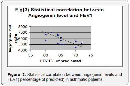
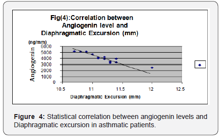
A significant negative correlation was found between
angiogenin levels and FEV1 (percentage of predicted) and
diaphragmatic excursion. In accordance with the above results,
Vrugt [22] and Salvato [18] showed that uncontrolled asthmatic
cases had more blood vessels in bronchial wall than patients
with controlled asthma due to higher levels of angiogenesis
factors, which suggests a possible relation between vascular
remodeling of the airway wall and asthma severity and control.
[19] concluded that patients with uncontrolled asthma display
chronic hyperinflation results from fixed airway obstruction
(airway remodeling) secondary to vasodilatation of blood vessels
in airways. This dynamic hyperinflation increases expiratory
flow limitation and impairs pulmonary functions. It also flattens
the diaphragm and reduces generation of force since muscle
contraction results from a mechanically disadvantageous fiber
length. Bronchial vascular remodeling, with an increase in size
and number of blood vessels as well as vascular hyperemia have
been proposed as contributing factors in airway wall remodeling
in patients with uncontrolled asthma [20].
This study showed that sEMG recording parameters,
including RMS of amplitude and frequency were significantly
decreased after MVV maneuver (decrease of 18% and 12% from
baseline, respectively, (P<0.05) in asthmatic children compared
to control. These results suggested that the diaphragm is
exposed to fatigue in asthmatics after the MVV maneuvers. This
was in agreement with other researchers [21,22]. A number
of mechanisms may be involved in decreased sEMG amplitude
and frequency of diaphragm in asthmatics. It was reported that
decrease of sEMG amplitude in the diaphragm might reflect
decreased motor unit recruitment due to decreased activation
of the target muscles caused by central fatigue [23] or decrease
of individual motor unit action potential [24]. The reduction
in median frequency of the EMG power spectrum has been
typically considered as an indicator of fatigue as it has been
noted during fatigue by maximum and sub-maximum voluntary
contractions [21,25]. The shift in the EMG power spectrum is
considered to be associated with fatigue-induced metabolite
accumulation, change in intracellular PH, and reduction in
muscle fiber conduction velocity. Changes in frequency could
also represent the change of the recruitment because previous
research reported that the frequency changed as the levels ofcontractions changes [24]. During quiet breathing, type I fibers
of diaphragm are predominately activated. Type II fibers are
required in conditions requiring more force generation as during
maximal voluntary breathing. Fatigue may result when type II
fibers can no longer be effectively activated to sustain the force
of contraction. Conventionally, the diaphragm is believed to be a
skeletal muscle that does not fatigue easily. Some investigators
suggested that MVV maneuvers might not cause the muscle itself
to fatigue, but induce the “protective” central fatigue. Because
peripheral muscle fatigue would result in persistent muscle
fatigue and longer recovery period, perhaps central nerve system
would induce an earlier fatigue to prevent further inspiratory
muscle fatigue [25].
This study explored the effect of 12 weeks of strengthening
exercise of diaphragm in asthmatic children. It revealed a
statistical significant higher values of FEV1, FEV1/FVC, PEF
and MVV and also a significant increase in diaphragmatic
thickness and excursion in asthmatic patients after diaphragm
strengthening exercise. Many authors studied the effect of non
respiratory exercise on pulmonary function parameters in
asthmatic children [19, 21,26-29,] Their results revealed marked
improvement in pulmonary functions after diaphragmatic
training. Girodo [29] studied the effect of 16-week program of
diaphragmatic strengthening exercise for asthmatic patients.
They found a significant reduction in medication use and in the
intensity of asthmatic symptoms. A follow up at two months
found that many patients have returned to earlier medication
levels with marked impairment in their PEF. McCool [30]
postulated that weight-bearing maneuvers may be used to
strengthen the diaphragm and expiratory muscles. However
they found that although strength training leads to myofiber
hypertrophy, it does not result in mitochondrial proliferation, so
weight lifting increases diaphragm structure and pressures with
less effect on contractility and excursion.
Al-Bilbesi and McCool [31] confirmed that during weightlifting
maneuvers, abdominal pressure is increased and the
diaphragm may be tensed to minimize the transmission of intraabdominal
pressure to the thorax. Consequently the diaphragm
is recruited and trans-diaphragmatic pressure is increased.
Weiner [21] studied the effect of specific diaphragm training
program in patients with asthma. They resulted in a significant
improvement in respiratory muscle performance and a decrease
in the sensation of breathlessness with decrease in β2-agonist
consumption. They explained that by the increased inspiratory
muscle strength in trained patients. Ramirez-Sarmiento [8]
observed an increase in both the strength and endurance of
diaphragm after diaphragm strengthening exercise with an
increase in proportion of type I fiber and decrease in type II
fibers. In addition both fiber types exhibited an increase in
cross-sectional area. They concluded that diaphragm training
induces a specific functional improvement of diaphragm and
adaptive changes in the structure.
DePalo et al. [27] measured diaphragm thickness by
ultrasound at baseline and 8 and 16 wk after diaphragm
strengthening exercise. After training, there was significant
increase in diaphragm thickness. They stated that the
diaphragm and abdominal muscles can be recruited during
diaphragm strengthening maneuvers. With these maneuvers,
trans-diaphragmatic pressures are elevated to levels that
could potentially provide a strength-training stimulus. Enright
[32] and Padula and Yeaw [33] suggested that high-intensity
inspiratory muscle training and diaphragm strengthening result
in increased contracted diaphragm thickness and increased
lung volumes and exercise capacity in patients with obstructive
airway diseases.
Conclusion
In future studies of respiratory muscle function in asthma
should be aided by measurement of diaphragm thickness and
excursion particularly in patients with severe hyperinflation
who are most likely to have impairment of muscle function.
Diaphragmatic training programs may be of value in improving
pulmonary functions of children with asthma.
sodium alendronate can be easily delivered through
inhalation devices. Dendrimers are getting attention as in these
days. These are nano-size synthetic macromolecules with a
highly branched structure and globular shape. Dendrimers
contain a large number of terminal groups, which allows these
molecules to bind with several drugs at the same time and can
deliver them with accuracy and precision.
There are several dendrimer-based formulations
available, such as anticancer drug fluorouracil attached to the
dendrimers with cyclic core and antitumor drugs adriamycin
and methotrexate with dendrimers having poly(ethylene
glycol) grafts. Precision and targeted drug delivery is rapidly
gaining importance now a days and considering the mechanical
improvement of formulation and delivery devices, nanoparticles
and nanoparticles-mediated drug delivery are at the peak.
However, since the characteristics of the engineered nanocarriers
or nano formulations are complex and may change in certain
solutions or devices, t is important to focus on the development
of appropriate devices to deliver the drugs effectively. Preclinical
and animal studies are extremely important at this stage to
investigate the efficacy and safety of these formulations before
bringing them as regular treatment modality.
To know more about Open Access International
Journal of Pulmonary & Respiratory Sciences please click on: https://juniperpublishers.com/ijoprs/index.php
To know more about Open access Journals
Publishers please click on : Juniper Publishers

Comments
Post a Comment